Complementary and Alternative Medicine Dissection Course 2015
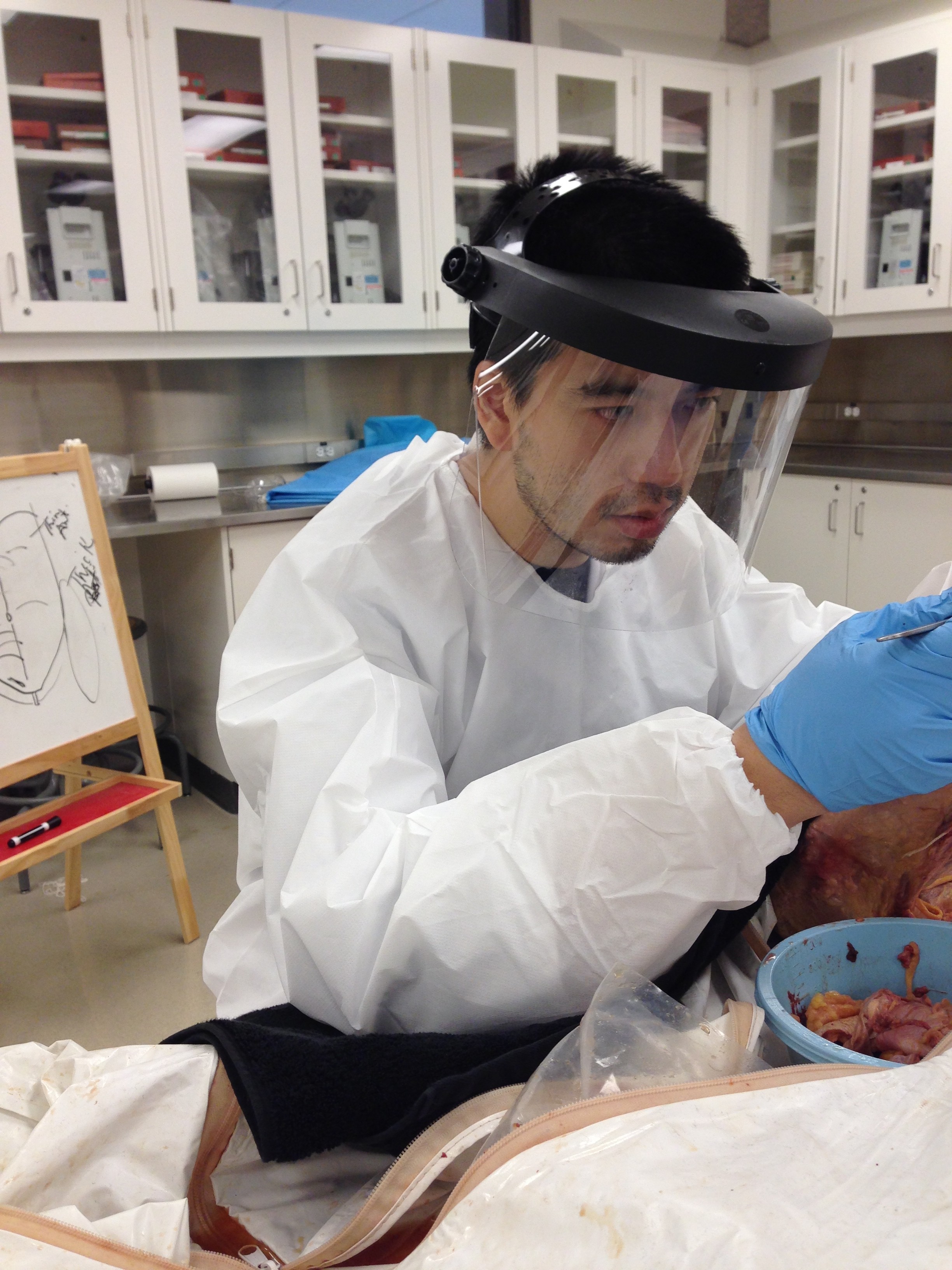
This summer, I dissected a 77-year old man. I’m contributing to a neuro anatomical textbook by using real human specimens.
It was in a summer course called the The Complementary and Alternative Medicine Human Dissection Experience (CAM – HD Experience). It’s at Queens University and it is run by Craig Harness MSc, DO(Q).
This is an incredible self-guided program where you get 10 days of dissection!
Dissection is an amazing experience for any health care practitioner because it allows you to see the body for what it’s really like – and not just what you see in textbooks. Textbooks show bodies as if we were “perfect” on the inside.
Without actually seeing a body “in the flesh” you can never really grasp how complex the body truly is.
Think about it… flying a plane is not like using a flight simulator, is it?
So what exactly is it like to dissect a human body?
Today I’m going to share with you everything I learned about human bodies after taking this dissection course.
Before I start, I should mention that the cadaver was donated in the name of science. He was a 77-year old man who passed away from cancer.
First, you should know that these “fully intact” bodies have been embalmed very lightly to preserve tissue consistency and joint range of motion. Embalming is the art and science of preserving the body and delaying bodily decomposition. This is what is done at a funeral, for example, so that the body stays suitable for public display. Embalming also preserves the joint range of motion – similar to raw chicken or beef.
This experience was entirely new to me because we had to remove the layers of the cadaver – starting with the skin.
All of the cadavers I have worked with in the past have been pre-cut and cleaned up to look like the textbooks. All the fat and loose connective tissue has been cut away so that the student experience is easy.
But this time, the cadaver was in its natural state. And cutting into each layer was like flipping through a book. Where you can turn each page (or muscle) and discover what is hidden underneath. And as you keep flipping the pages, you discover what is underneath every layer.
This is me after finding the Dorsal Scapular nerve and Artery! I went through 5 shoulders finding this nerve!
The very first layer we started out with was the epidermis. Then we moved on to the adipose tissue, which is our body fat. Then we moved on to each section of muscle we could identify.
As we peeled away the layers, we found golf ball like nodules throughout the armpit and groin area suggesting that he passed away from some form of lymphoma. We cut one out and sliced it into thin layers to see the consistency of this cancer. It looked like a dense processed meatball.
On another body we found thyroid cancer – it was the size of a huge peach! When we cut into it, it looked like pomegranate on the inside with clusters of red nodules.
Are you still with me?
Good!
After uncovering these bodies layer by layer, here’s what I learned:
#1 Your muscles and organs are extremely protected by fascia.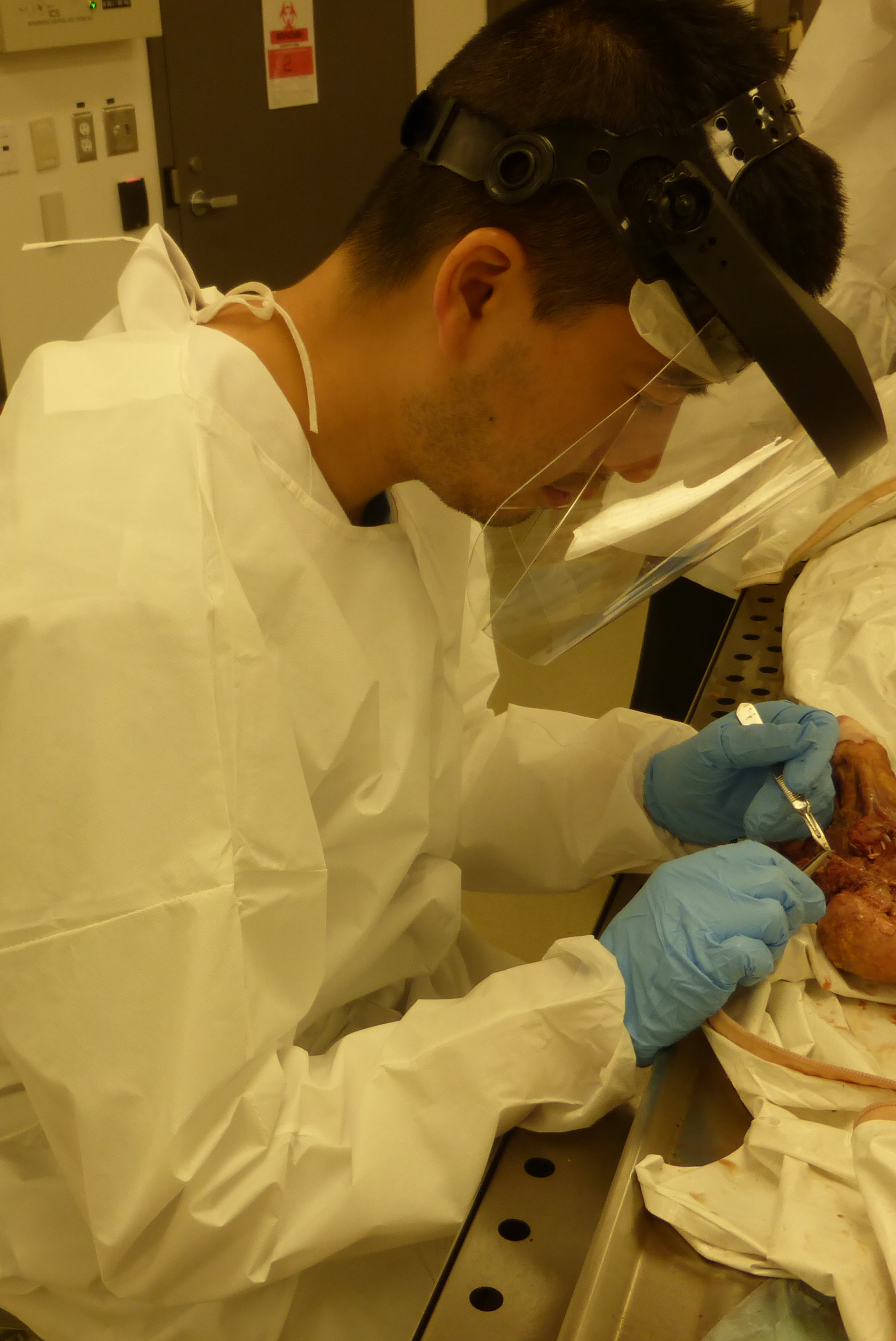
As I’m pulling back the layers of tissue, there is a “cotton candy” substance between the layers called fascia. This is the tissue that is “cleaned up” with a scalpel. The fascia takes up space between the layers of tissue, but this fascia envelopes and binds all tissues together to form a working unit. Fascia grows in layers around muscles and organs. In some areas it is difficult to determine where some structures end and begin.
All tissues are embedded together in some form of fascia.
I could not really appreciate fascia until now. Textbooks are very much watered down. They have pictures of very clean “perfectly dried” specimens, when in fact, it is much more complex!
#2 Fat is EVERYWHERE…
If there’s a space in the body, fat will occupy that space. The fat is used to protect sensitive areas. Sensitive areas are places where nerves cluster or the areas around important blood vessels.
The fat that protects the kidneys is thick! In textbooks, the fat is all removed and cleaned up for learning purposes.
Ever wondered what nerves feel like?
#3 Nerves feel like seatbelts.
When you pull on the nerves, there is very little give – yet it is flexible. Seeing the nerves made me really appreciate how neural flossing can help. Neural flossing is where specific exercises are used to improve mobility of different tracts of the nervous system.
When you get acupuncture, the needle is intended to target the point where nerves come out of structures or divide. These fine nerves need to be cleaned so that structures exposed are identifiable for textbook quality print.
#4 Cracking a human skull with a hammer IS much harder than Hollywood makes it look!
You need to hit the skull just enough so that the thinner parts of the bone can come off easily (after you have sawed it). Once the skull is cracked, we have to quickly go through each layer of the brain because the moment we took off the outer lining of the brain (the Dura) the brain itself began to oxidate and melt. Removing the layers of the brain was truly a unique experience in and of itself.
One of the most disturbing feelings was sawing through bone with a hacksaw and bandsaw. It almost felt like I was killing a live being. Another unique and disturbing feeling was dislocating a hip joint.
Overall, it was an incredible experience that I would HIGHLY recommend to any health care practitioner.
Fair warning: the process was extremely slow because you have to manually trim away a TON of fascia and adipose tissue. The good news is, as soon as you cut it out – it’s gone! Imagine carving out a book from a tall, mature tree using only a scalpel and a pair of tweezers….that’s what this was like!
Beyond the pure love for science and the human body, I was taking this course to assist Dr. Poney Chiang with the Acupuncture textbook. This course allows me to expose the critical nerve structures for acupuncture. Stay tuned for more exciting developments on this acupuncture publication!
As for the dissection course, I plan to be there again for 2016 and hope to see you there with level 2 biohazard suits on (no bells or open toed shoes please).
I just wish to add a big thank you to our body, and his family. Without your consent to donate to science, we would not have had this great opportunity to understand the human body.
If you have any questions, please don’t hesitate to ask me, Samuel Lo at the Toronto Acupuncture Clinic!
As always, thanks for reading and sharing with your colleagues, friends and family.
Samuel Lo
incredible and fascinating- great to know how our body is layered.
Thank you very much for sharing your experience of dissecting a human. It is awesome and very interesting to know the formation of our body., the tissues, the organs how they are protected etc. I am sure you had an excellent course to know more about human bodies by dissecting one on your own. I also took a course from you and find it very interesting. Well done!
I loved reading your blog! it was amazing! i can’t believe you actually cut up your own body! its like breaking bad, but for science! Keep up the great work! i look forward to reading your next blog! SAM THE MAN!
Neat. Confirms what my body tells me about it being all connected! Now, how does one TREAT fascia issues, as opposed to it being something you just scrape away? This illuminates the next direction for health treatment, and the understanding of Chinese methods.